Wound Image Analysis
Management of chronic illnesses, such as Type 2 diabetics, is challenging for both patients and the health care system. Due to poor circulation, often develop chronic wounds that are slow to heal and that require daily care. Given the cost and inconvenience of regular visits to wound clinics, we are developing image analysis tools that can complement to services rendered by wound clinics. Specifically, we ware developing a smart phone app (Android), which tracks the wound size and wound healing process based on images captured with a smart phone. But weight management, exercise, diet and glucose monitoring are also important in order for the wound to heal properly, and thus the smart phone app is set up stop also acquire such data and to provide constructive feedback.
The wound image is captured by the smart phone camera and transmitted to an embedded PC via Wi-Fi for image processing. The PC decompresses the image and converts it to bitmap format, then segments the wound in the image by employing the distance regularized level set evolution (DRLSE) method, which has the advantages of the level set algorithms while eliminating the need for re-initialization of level set function. Next, color segmentation within wound boundaries is performed by applying the K-Mean color clustering algorithm based on red-yellow-black (RYB) evaluation model for wound healing. Finally, the results are re-formatted, transmitted back to the smart phone and displayed.
To accelerate the segmentation, we implement the DRLSE method on the GPU & CPU cooperative hardware platform in data-parallel mode, which has improved the computational efficiency. Processing wound images acquired from UMASS Medical Center has demonstrated that the wound image analysis system provides accurate wounds area determination and color segmentation. For all wound images of size of 576*324, with complicated wound boundaries, the wound analysis consumed max 2 seconds, which is 5 times faster than the same algorithm running on CPU alone.
To evaluate the performance of wound image processing algorithms, we implemented the entire method shown in the figure below on sample wound images obtained from patients at UMASS Medical Center, without any attributing information. The processing results are promising and the processing time is at most 2 seconds, which is sufficient for a real time image processing system. As shown in the image sequence below, (a) is the original wound image displayed on the screen of a Motorola smart phone, and (b) is the wound image with boundaries indicated. In (c), we can see that the different color tissues (here only red and yellow) are successfully separated by K-mean clustering algorithm.
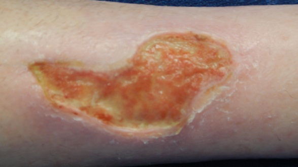
(a) Original wound image
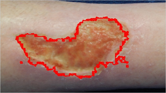
(b) Wound boundary determination
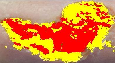
(c) Wound color segmentation